


Vascular Occlusions
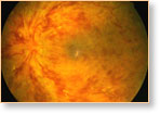
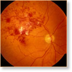
A retinal vein occlusion (vascular occlusion) occurs when a blockage in the vein prevents adequate blood flowing into the affected area of the eye. There are two types of this kind of problem — central retinal vein occlusion (CRVO) or branch retinal vein occlusion (BRVO).
Understandably, the central form, which affects the main retinal vein, is the more serious, while the branch form relates to a vein blockage in one specific part of the eye.
These problems are more likely to occur among people who already suffer from:
- Glaucoma
- Diabetes
- Age-related blood vessel problems
- High blood pressure
- Blood disorders
The symptoms of retinal vein occlusion include blurred vision, floaters and, at times, pain in the eye. The blurred vision occurs when blood, leaking from the blocked vein, collects in the macula — the centre of the retina. As described in the floaters section of the website, floaters can appear as spots or other shapes, interfering with normal vision. These are usually caused by the leaking of blood into the vitreous, causing shadows to be cast on the retina. When pain occurs, it is usually related to excessive eye pressure called neovascular glaucoma. It is a complication created when the central retinal vein is involved.
While surgery is not always possible for retinal vein occlusions, it is possible for laser surgery to be effective for some patients suffering from branch retinal vein occlusion, which has generated macular edema.
When surgery is possible, it is strongly recommended that it take place as early as possible following diagnosis.